Our Technology
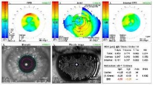 OPD SCAN III
The OPD Scan III is used during each comprehensive examination. This scan combines three advanced technologies in addition to a detailed corneal analysis. One scan gives our doctors diagnostic information on the internal structures of the eye and includes a topography of the cornea as well as an approximate refraction. Each component of the OPD scan insures a detailed diagnosis that in the past was simply unavailable.
 OPTOMAP DIGITAL IMAGING
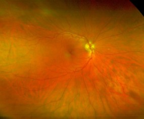
We believe the Optomap Retinal Exam is a vital part of every examination. Optomap is painless and the only technology that provides an ultra-wide view of the retina without the need for dilation drops. This complete view enable our doctors to more easily detect diabetes, high blood pressure, macular degeneration, glaucoma and other eye health issues.
After reviewing the images with your doctor, each patient is able to make informed decisions about their eye health and wellness. The images are archived and used to monitor your eye health from year to year.
 ZEISS OCT DIGITAL SCAN
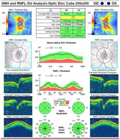
At Palm Beach Eyes we have always believed in the need to stay abreast of the latest developments in technology and know that it is through clinical distinction that patients visit our practice. We are one of the few practices in Palm Beach County to have invested in high resolution OCT (Optical Coherence Tomography) technology which allows us to offer a unique service to patients.
OCT technology is the next generation in high-resolution imaging of the retina and other internal structures of the eye, effectively an “optical ultrasound”. The unique way in which the image is formed means that for the first time it is possible to see below the surface of the retina and view the microscopic layers beneath. This is of profound importance in determining the precise diagnosis of visual problems and helping guide patients with their treatment options. Everyone would benefit from an OCT scan.
The scanning procedure is painless, noninvasive and is similar to having a photograph taken, but with the camera positioned close to the eye. Nothing touches the eye and there is no discomfort during the scan. OCT scans are like viewing a biopsy of the retina on a high powered microscope without ever touching the eye. These scans can also be put together to produce a 3D image of the back of the eye, which is a very powerful tool to show small detail and changes that could cause future problems.
 ZEISS VISUAL FIELD
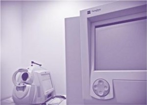
Visual field testing measures both central and peripheral vision and visual function. A visual field test provides vital information used to diagnose and monitor many eye or neurologic problems and documents vision loss or obstruction. The computerized perimeters of the test provide a comprehensive report and picture of the visual field along with any defects in the field. A visual field test is extremely valuable because it is a noninvasive, rapid method of gathering a wealth of information regarding a person’s ocular and neurological health. Visual field testing is the only way to document actual visual loss and whether the loss is progressing or remaining stable.